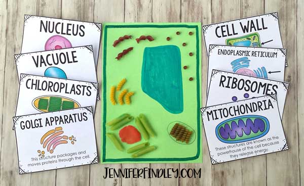
23+ Animal Cell Project Ideas
Edible cell models can be eaten yum and are often made with cake large cookies Rice Krispie Treats Jell-O berries or candies eg MMs gummy worms jelly beans etc. Make thin line shape of yellow color play dough for golgi bodies.

Best 25 Animal Cell Project Ideas On Pinterest Cell Cute766
Its no secret that people choose unique ideas certainlyfor memorable moment - at this siteare 10 artistic 3D Animal Cell Project Ideas.
Animal cell project ideas. 10 fascinating 3D Animal Cell Project Ideas so that anyone will never have to explore any further. 13 cup vegetable oil. Sep 19 2015 - Explore Jeremiah Johnsons board animal cell project on Pinterest.
The following animal project ideas help to introduce animal behavioral study in many different species. People also love these ideas. Carefully shape it as semi sphere shape.
This is cell membrane of our 3d animal cell. Non-edible cell models cannot be eaten and are often made with everyday craft supplies like styrofoam pipe cleaners shower gel string Play-Doh or modeling clay. See more ideas about Cells project Animal cell project Animal cell.
Pull apart the Pull n Peel Twizzlers to separate the orange and red vines. Once your cake batter is smooth add a few drops of food coloring to mimic the pink color of many animal cells cytoplasms. First of all take a big chunk of blue color play dough.
Searching for a special ideas has certainly never been easier. They are nucleus and nucleolus. Feb 27 2018 - Save this one for the Science Fair.
See more ideas about cells project plant and animal cells cell model. 3d Animal Cell Project Edible Cell Project Cell Model Project Cell Project Ideas 3d Plant Cell Model 3d Cell Model 3d Animal Cell Model Biology Projects School Projects. Nov 7 2015 printable animal cell diagram to help you learn the organelles in an animal cell in preparation for your test or quiz.
May 1 2018 - Explore Shannon Browns board Animal cell project on Pinterest. Nov 13 2016 - Animal Cell Model More. The outer chocolate layer represents the nucleus and the creamy peanut butter center acts as the nucleolus.
Large and filled with fluid storage tanks for cell waste. The gelatin in your cell model represents cytosol. How to DIY a 3-D Model of an Animal Cell.
Next place a peanut butter cup in the middle of the cell. Gumball or other larger spherical candy. See more ideas about cells project animal cell project animal cell.
Your 3d animal cell model may not look like typical cell diagrams but actually building a 3D model will help you visualize and remember the parts of the cell. When your cake batter is an even color pour it into a square cake pan and bake it in the oven at 350 degrees for about 30 minutes. Be sure to get permission from your instructor before beginning any animal science projects or behavioral experiments as some science fairs prohibit these.
Sep 22 2020 - Explore Debi Liggetts board Project ideas on Pinterest. Animal Cell Handout Animal Cell Project Animal Cells Model Animal Cell 3d animal cell project plant cell project cell model project human cell diagram science projects. After this take orange color and yellow color play dough and place at middle.
The Red Vine border represents the cell wall.
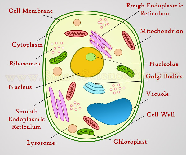
51 Animal Cell Organelles Diagram
Learn vocabulary terms and more with flashcards games and other study tools. Animal cell organelles labeled diagram.

The Nucleus And Cytoplasm Anatomy And Physiology
Cell organelle is a specialized entity present inside a particular type of cell that performs a specific function.
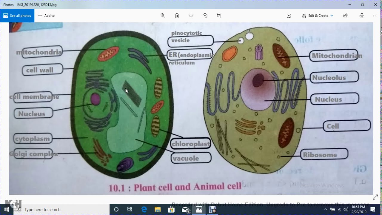
Animal cell organelles diagram. Start studying Animal Cell Organelles Diagram. Eukaryotic cells are larger more complex and have evolved more recently than prokaryotes. Cell Organelles definition.
Selectively permeable structure that controls what substances come into and out of a cell. It is mainly made up of water and protein material. The structure of an animal cell differs slightly from a plant cell in terms of shape protective covering and organelles.
Listed below are the Cell Organelles of an animal cell along with their functions. 9 sets of 3 microtubules that are important in cell. There are two types of cells - Prokaryotic and Eucaryotic.
A labeled diagram of the animal cell and its organelles there are two types of cells prokaryotic and eucaryotic. In the labeled animal cell diagram it is nearly circular in shape and lacks outer cell wall. Identify the organelles that are numbered and the function of each organelle by.
Where prokaryotes are just bacteria and archaea eukaryotes are literally everything else. The various cell organelles present in an animal cell are clearly marked in the animal cell diagram provided below. Major cell organelles are as follows.
Start studying Animal Cell Organelles. Animal cell diagram detailing the various organelles Though this animal cell diagram is not representative of any one particular type of cell it provides insight into the primary organelles and the intricate internal structure of most animal cells. Rod-shaped structure that converts energy in food to energy the cell can use.
Its the fluid that contains the organelles. There are various cell organelles out if which some are common in most types of cells like cell membranes nucleus and cytoplasm. Learn vocabulary terms and more with flashcards games and other study tools.
The cell organelles found in the animal cell are plasma membrane centriole peroxisome lysosome ribosomes mitochondria endoplasmic reticulum cytoplasm nucleus nucleolus nuclear envelope and golgi apparatus. There are various organelles present within the cell and are classified into three categories based on the presence or absence of membrane. 11 Cytosol Its not an organelle.
Learn vocabulary terms and more with flashcards games and other study tools. Start studying Animal cells organelles. The cell membrane is the outer most part of the cell which encloses all the other cell organelles.
8 Smooth endoplasmic reticulum SER. While the plant cell resembles rectangular shape and possesses a rigid cell wall. Differences in cellular structure of prokaryotes and eukaryotes include the presence of mitochondria and chloroplasts the cell wall and the structure of chromosomal DNA.
An animal cell structure is very complex from other organisms except for plants because there are many organelles present inside the cell of an animal cell. Eukaryotic cells contain membrane-bound organelles such as the nucleus while prokaryotic cells do not. Well-Labelled Diagram of Animal Cell The Cell Organelles are membrane-bound present within the cells.
In short the outer layer of an animal cell is the flexible membrane. Animal cell structure animal cells have a variety of different organelles that work together to allow the cell to perform its functions. Learn vocabulary terms and more with flashcards games and other study tools.
Centrioles are about 500nm long and 200nm in width that are. 5 Rough endoplasmic reticulum RER. A Labeled Diagram of the Animal Cell and its Organelles.
In the labeled animal cell diagram it is nearly circular in shape and lacks outer cell wall. The cell membrane controls the influx of the nutrients and minerals in and out of the cell.
84+ Animal Cell Parts 7th Grade
Develop and use a model to describe the function of a cell as a whole and ways parts of cells contribute to the function. You can edit this venn diagram using creately diagramming tool and include in your reportpresentationwebsite.

Pin By Az Always Creations On For My Kids Animal Cell Anatomy Cell Parts Animal Cells Model
Study of animal cell helps us to understand about whole body.
Animal cell parts 7th grade. 16 rows cell membrane. 7 Class or period 1 2 3 4 5 6 7 8. Lesson Plans for the Animal Cell.
Endoplasmic reticulum ER PlantAnimal A network of passages that. Descubre y guarda tus propios Pines en Pinterest. Sub strand - A.
Most commonly found in plant cells. Packages them and distributes them to other parts of the cell. 7th Grade Life Science.
As well as animal cells obtained using FEM so that color is not the rule. Plant and animal cells have parts called organelles that help them function and stay organized. The student will understand that all organisms are composed of cells that carry.
Use this page to help you complete the Cell Jobs worksheet. Animals and Scientfic Inquiry. Both animal and plant cell.
A Cell is the basic unit of life. 7th Grade Science Understanding Our Cells. 1 2 3 4 7 8 13 12 11 10 9 5 6 14.
Cell membrane is outer most part of animal cell. We will cover the nucleus cell wall cell membrane mitochondria chloroplast lysosomes and vacuoles. 7th Grade Science - Plant and Animal Cell Vocab.
7th grade plant and animal cell venn diagram. Plant and Animal Cell Vocabulary. Improve your science knowledge with free questions in Identify functions of animal cell parts and thousands of other science skills.
Cell Parts - simple PowerPoint show comparing organelles of plant and animal cells. 18-nov-2013 - Norma Denham descrubrió este Pin. Animal cells with diagrams animal cell srah unverzagt cell models 2018 culmination.
Plant and animal cells. Developing and using models. Students will be asked to compare and contrast animal and plant cells.
7th Grade Science. Cell Theory and Cell Functions Grade. Minnesota Science Standard.
Outer membrane of cell that controls movement in and out of the cell Double layer. A protective outer covering - regulates interaction between the cell and its. Grade 7 - Cells Functions of Organels.
Clink on the name of the cell part to find out. Learn vocabulary terms and more with flashcards games and other study tools. Virtual Cell - roll your cursor over the cell drawing.
Desired Results Established Goals Standards SCI712C Recognize levels of organization in plants and animals including cells tissues organs organ systems and organisms. Comparison of Plant and Animal Cells - 26 slides including a Venn diagram for comparing. There are may parts inside a animal cell.
Plant and Animal Cells. 25 plant cell diagram 7th grade. On the many functions needed to sustain life.
The Animal Cell. This rap was created for a 6th-grade science classroom to teach about the different parts of a cell. With its catchy rhythm and rhymes students of all learn.
You may need to edit it before using in 7th grade science. SCI712D Differentiate between structure and function in plant and animal cell organelles including cell. Golgi body PlantAnimal Receives materials form the ER packages them and sends them to other parts of the cell.
Both animal and plant cell. 7th Science Stage 1. Jan 24 2020 plant cell diagram 7th grade.
Plant and animal cells. Terms in this set 14 Cell wall. Plant and Animal Cells.
They will also need to be able to identify cell organelles by appearance and function. They are cell membrane nucleus nucleolus nuclear membrane cytoplasm endoplasmic reticulum Golgi apparatus ribosomes mitochondria centrioles vacuoles etc. What you will learn from this video Cells are the basic unit of all living things.
1 2 3 7 13 12 11 10 9 8 4 5 6. Mitochondria PlantAnimal Produces much of the cells energy. Proteins and other newly formed materials from the endoplasmic reticulum packages them and distributes them to other parts of the cell.
7th grade cell parts and functions. It performs all the the Life Functions.
36 Animal Vacuole Cell
In plant cells vacuoles help maintain water balance. They arent needed as much for breaking down substances because lysosomes another organelle in animal cells do that.
Plants Vs Animals Bronco Biology Exploring The Cell
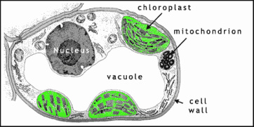
Made of a tough substance called cellulose which supports the cell.

Animal vacuole cell. They are found in both animal and plant cells but are much larger in plant cells. Especially in protozoa single-celled eukaryotic organisms vacuoles are essential cytoplasmic organs organelles performing functions such as storage ingestion digestion excretion and. The vacuoles of the animal cells are useful for overcoming the foreign particles that may be the bacteria.
Plant and animal cell diagrams quiz. Vacuolesare storage bubbles found in cells. Initially the vacuole is like a small bubble.
Vacuoles in animal cells. It is the obligation of the membrane of the cell to invaginate for the prime goal of engulfing the bacteria. Plant cells also have a cell wall and often have chloroplasts and a permanent vacuole.
Vacuole Function in Animal Cells. The vacuole is a type of organelle present in eukaryotic cells. Vacuoles might store food or any variety of nutrients a cell might need to survive.
Sometimes a single vacuole can take up most of the interior space of the plant cell. Plant cells consist of a cell wall which helps to protect and support the cell. This enhanced visual instructional tool assists in grasping and retaining the names of the cell parts like mitochondrion vacuole.
In this process a vacuole is formed. Some animal cells do not have vacuoles. Vacuole in biology a space within a cell that is empty of cytoplasm lined with a membrane and filled with fluid.
However some protists animal cells and bacteria also contain vacuoles. In animal cells vacuoles are generally small and help sequester waste products. Contains a liquid called cell sap which keeps the cell firm.
It contain fluid called cell SAP which consists of water sugar amino acids in Science salt nitrogenous waste etc. It becomes larger as the cell grows. In animal cells they are small and typically transport materials into and out of the cell.
In animal cells vacuoles tend to play a lesser role. Animal cell vacuoles are much smaller than plant or fungal vacuoles and animal cells generally have multiple vacuoles. Vacuoles can store different substances depending on the type of cell they are in.
In animal cells vacuoles perform a more subordinate role as mediating storage units and carriers during exocytosis and endocytosis. It is a sac surrounded by a single membrane called a tonoplast. Animal cells usually have an irregular shape and plant cells usually have a regular shape.
Vacuoles are fluid-filled enclosed structures that are separated from the cytoplasm by a single membrane. Even though plant cells and animal cells both have vacuoles the vacuole present in the plant cell is much larger compared to the one in the animal cell. For example in fat cells vacuoles will often store large amounts of lipids.
The plant vacuole stores water whereas animal vacuole store nutrients ions waste products and water The plant vacuole is located at the center of the cell while animal vacuole is distributed all over the cell. A vacuole is a membrane-bound cell organelle. The animal vacuole is suitable for exocytosis and endocytosis whereas plant cell is responsible for maintaining turgor pressure.
They can even store waste products so the rest of the cell is protected from contamination. Plant and animal cells. The main function of vacuoles in animal cells is to isolate and remove waste products from.
Animal cell vacuoles are typically small and each cell can contain multiple vacuoles. Vacuoles in animal cells mostly store substances. Vacuoles serve many functions depending on the needs of the cell.
They are found mostly in plant cells and fungi. A vacuole is an organelle inside plant and animal cells that stores water and some wasteAn animal has small vacuoles which are barely more than large vesicl. Vacuoles are storage sacs or cavities in which solid or liquid is stored in the cell.
Although animal cells contain vacuoles they do not contain large central vacuoles. A vacuole is a cell organelle found in a number of different cell types.
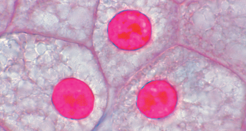
35+ Animal Cell Under Microscope
Similarly what does a animal cell look like under a microscope. Viewing animal cells under the microscope and calculating magnification.
Slide Animal Cell Sec
Likewise can rough endoplasmic reticulum be seen under a light microscope.
Animal cell under microscope. Under a microscope plant cells from the same source will have a uniform size and shape. Thousands of new high-quality pictures added every day. Animal cells under microscope animal cells under microscope labeled animal cells under microscope video animal cell under microscope 400x animal cell under microscope 40x animal cell under microscope 100x animal cell under microscope 10x animal cell.
Human cheek cells are made of simple squamous epithelial cells which are flat cells with a round visible nucleus that cover the inside lining of the cheekC. Under a microscope plant cells from the same source will have a uniform size and shape. These cell organelles perform specific functions within the cell.
The Cell as Seen under the Electron Microscope. Some of the cell organelles that can be observed under the light microscope include the cell wall cell membrane cytoplasm nucleus vacuole and chloroplasts. Section of beef marrow showing motor neurons in the anterior horn seen under a microscope.
Browse 163 animal cells under microscope stock photos and images available or start a new search to explore more stock photos and images. An animal cell also contains a cell membrane to keep all the organelles and cytoplasm contained but it lacks a cell. Microscopically animal cells from the same tissue of an animal will have varied sizes and shapes due to the lack of a rigid cell wall.
Observing Animal Plant Cells You will be observing plant and animal cells under the microscope. Scientists work hard to make accurate observations. Beneath a plant cells cell wall is a cell membrane.
To look at a cell close up we need a microscope. Plant cell as shown above. The shape of both cells were easily seen and some similarities and differences were.
Under the microscope animal cells appear different based on the type of the cell. Animal cell under the microscope A typical animal cell is 1020 μm in diameter which is about one-fifth the size of the smallest particle visible to the naked eye. Microscope is used extensively in cell biology microbiology biotechnology microelectronics nanophysics pharmacology mineralogy and forensics.
Cells are the smallest part of a living organism and are around 001 mm - 003 mm long. Beneath a plant cells cell wall is a cell membrane. In a plant cell the nucleus cell wall cell membrane and the cytoplasm were visible through a light microscope.
Diagrams from enchanted learning. The lesson will also take you through some exam questions on finding magnification using a scale and an image of a cell. The cell membrane is the outer most part of the cell which.
It is also used for medical diagnosis particularly while dealing with tissues or in smear tests on free cells or tissue fragments. Careful observations help scientists make predictions draw conclusions and find comparisons. Find animal cell under microscope stock images in HD and millions of other royalty-free stock photos illustrations and vectors in the Shutterstock collection.
Start studying Cells Under a Microscope Animal Cell Plant Cells. If a plant cell and an animal cell are observed under a microscope what are the characteristics of the cells that enable you to identify the cell as a plant cells. Animal Cell as shown above.
Viewing Animal Cells under a microscope. It is very important that you take the time to make careful observations when looking at cells. Find out how to observe cells under a microscope Click to.
In the animal cell the nucleus cell membrane and cytoplasm were visible through a light microscope. Learn vocabulary terms and more with flashcards games and other study tools. In this lesson you will learn the method of how to view an animal cell under the microscope.
Cell nuclei of a sea urchin egg Parechinidae seen under a microscope. In plant cells but not animal cells you will find i a cell wall ii green chloroplasts and iii vacuoles. Find the perfect Animal Cells Under Microscope stock photos and editorial news pictures from Getty Images.
Select from premium Animal Cells Under Microscope of the highest quality.
