37 Animal Cell Under Electron Microscope
We all do not forget that the human physique is quite problematic and a method I. The cell membrane also known as plasma membrane or plasmalemma consists of three layers when viewed under the electron microscope.

Y4uq Xpinhgurm
Microscopically animal cells from the same tissue of an animal will have varied sizes and shapes due to the lack of a rigid cell wall.
Animal cell under electron microscope. Animal Cell as shown above. IB Biology HLSLA Worksheet Microscopes and Electron micrographs Prokaryotic and Eukaryotic cells. It has small vacuoles.
6 rows Structure of plant and animal cells under an electron microscope. Some of the cell organelles that can be observed under the light microscope include the cell wall cell membrane cytoplasm nucleus vacuole and chloroplasts. It is an electron micrograph of cells largest and most important organelle the mitochondria and is characterized by the following features Fig.
The Cell as Seen under the Electron Microscope. Diagram Of Animal Cell Under Electron Microscope Labeled. Table D leads to images of electron microscopes or protocols for tissue preparation.
1 The name mitochondria was given by Benda 1898 and their ma n function was brought to light by Kingsbury 1912. Monday April 5th 2021. This atlas offers informative texts on a lot of cell organelles and ultrastructures with detailed information in a generally intelligible way.
It is completed by a vocabulary of microscopic anatomy. In the given figure of an animal cell as observed under an electron microscope. So lets begin by drawing a rough-oval shape.
2 Each mitochondria in section appears as sausage or cup or bowl shaped structure lined by double. Also know what does a animal cell look like under a microscope. But at the same time it is interpretive.
The three layers are composed of one layer of phospholipid sandwiched between two protein layers. The cell membrane is. Animal cells have a basic structure.
View under scanning electron microscope yeast cells of the. Under the intense radiation of the electron microscope 011 electron per Å 2 the question of viability of cells naturally arises because the amount of radiation absorbed during highmagnification imaging is sufficient to cause cell death. Typical Animal Cell With Labels Removed 3.
The ultrastructure of budding and dimorphic yeast cells observed with a scanning electron microscope sem and a transmission electron microscope tem after thin sectioning and freeze etching are then described followed by discussion of the regeneration of the cell wall ofcandida albicans. Below the basic structure is shown in the same animal cell on the left viewed with the light microscope and on the right with the transmission electron. Beneath a plant cells cell wall is a cell membrane.
Use your ebook to answer the questions below. Plant cell as shown above. Diagram Of Animal Cell Under Electron Microscope.
Most cells both animal and plant range in size between 1 and 100 micrometers and are thus visible only with the aid of a microscope. How is it different from animal cell. Asked Nov 28 2017 in Class IX Science by ashu Premium 930 points.
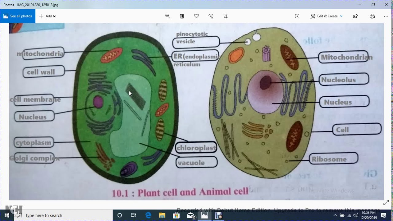
Typical Animal Cell Pinocytotic vesicle Lysosome Golgi vesicles Golgi vesicles rough ER endoplasmic reticulum Smooth ER no ribosomes Cell plasma membrane Mitochondrion Golgi apparatus Nucleolus Nucleus Centrioles 2 Each composed of 9 microtubule triplets Microtubules Cytoplasm Ribosome 2. Its a thin slice. These cell organelles perform specific functions within the cell.
Heres a diagram of a plant cell. The diagram is very clear and labeled. Summarize two advantages and disadvantages of light microscopes.
Structure of plant and animal. Human cheek cells are made of simple squamous epithelial cells which are flat cells with a round visible nucleus that cover the inside lining of the cheekC. Please show the substitute teacher your completed work as soon as you finish.
Illustrate only a plant cell as seen under electron microscope. However no obvious structural damage was apparent and several repeated scans gave the same images. Under a microscope plant cells from the same source will have a uniform size and shape.
It is flexible and has pores. Cell Structure and Function Student Hadi Yaafar Date May 24 2021 Instructions Please work independently. This feature was lost in the distant past by the single-celled organisms that gave rise to the kingdom Animalia.
Unlike the eukaryotic cells of plants and fungi animal cells do not have a cell wall. I Name the parts labelled as 1 to 10.
51 Animal Cell Organelles Diagram
Learn vocabulary terms and more with flashcards games and other study tools. Animal cell organelles labeled diagram.

The Nucleus And Cytoplasm Anatomy And Physiology
Cell organelle is a specialized entity present inside a particular type of cell that performs a specific function.
Animal cell organelles diagram. Start studying Animal Cell Organelles Diagram. Eukaryotic cells are larger more complex and have evolved more recently than prokaryotes. Cell Organelles definition.
Selectively permeable structure that controls what substances come into and out of a cell. It is mainly made up of water and protein material. The structure of an animal cell differs slightly from a plant cell in terms of shape protective covering and organelles.
Listed below are the Cell Organelles of an animal cell along with their functions. 9 sets of 3 microtubules that are important in cell. There are two types of cells - Prokaryotic and Eucaryotic.
A labeled diagram of the animal cell and its organelles there are two types of cells prokaryotic and eucaryotic. In the labeled animal cell diagram it is nearly circular in shape and lacks outer cell wall. Identify the organelles that are numbered and the function of each organelle by.
Where prokaryotes are just bacteria and archaea eukaryotes are literally everything else. The various cell organelles present in an animal cell are clearly marked in the animal cell diagram provided below. Major cell organelles are as follows.
Start studying Animal Cell Organelles. Animal cell diagram detailing the various organelles Though this animal cell diagram is not representative of any one particular type of cell it provides insight into the primary organelles and the intricate internal structure of most animal cells. Rod-shaped structure that converts energy in food to energy the cell can use.
Its the fluid that contains the organelles. There are various cell organelles out if which some are common in most types of cells like cell membranes nucleus and cytoplasm. Learn vocabulary terms and more with flashcards games and other study tools.
The cell organelles found in the animal cell are plasma membrane centriole peroxisome lysosome ribosomes mitochondria endoplasmic reticulum cytoplasm nucleus nucleolus nuclear envelope and golgi apparatus. There are various organelles present within the cell and are classified into three categories based on the presence or absence of membrane. 11 Cytosol Its not an organelle.
Learn vocabulary terms and more with flashcards games and other study tools. Start studying Animal cells organelles. The cell membrane is the outer most part of the cell which encloses all the other cell organelles.
8 Smooth endoplasmic reticulum SER. While the plant cell resembles rectangular shape and possesses a rigid cell wall. Differences in cellular structure of prokaryotes and eukaryotes include the presence of mitochondria and chloroplasts the cell wall and the structure of chromosomal DNA.
An animal cell structure is very complex from other organisms except for plants because there are many organelles present inside the cell of an animal cell. Eukaryotic cells contain membrane-bound organelles such as the nucleus while prokaryotic cells do not. Well-Labelled Diagram of Animal Cell The Cell Organelles are membrane-bound present within the cells.
In short the outer layer of an animal cell is the flexible membrane. Animal cell structure animal cells have a variety of different organelles that work together to allow the cell to perform its functions. Learn vocabulary terms and more with flashcards games and other study tools.
Centrioles are about 500nm long and 200nm in width that are. 5 Rough endoplasmic reticulum RER. A Labeled Diagram of the Animal Cell and its Organelles.
In the labeled animal cell diagram it is nearly circular in shape and lacks outer cell wall. The cell membrane controls the influx of the nutrients and minerals in and out of the cell.
27+ Animal Cell Anatomy And Physiology
1 Cells basic unit of life that is the building block of everything -makes up all tissues of an organism-performs all the functions by which life is defined including growth reproduction and metabolism 2 Tissues many cells grouped together that work to increase the speed efficiency and ability with which cellular tasks are performed. This is where respiration happens.
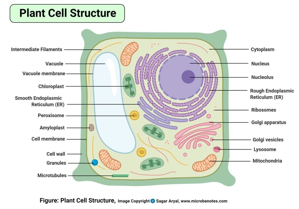
Plant Cell Definition Labeled Diagram Structure Parts Organelles
Animal anatomy snake diagram 6 photos of the animal.
Animal cell anatomy and physiology. A cell is the basic structural and functional b. A part of the cell containing DNA and RNA and responsible for. Even at the outset in the cell chapter lots of information that is lacking on cell function.
ANS 201 ANATOMY AND. A tubular organelle composed of nine triplets of microtubules that aids in the process of cell division. Cells Tissues Animal Welfare Show Class Anatomy and physiology of Domesticated Animals.
Centrioles split in two and migrate to opposite poles of a dividing cell to organize the spindle fibers enabling the cell to divide in two. 53 Cards 3 Decks 4 Learners Sample Decks. Chris Murungweni 22 Oct 2012 The overall objective is help students develop.
Learn anatomy and physiology animal cells with free interactive flashcards. A good knowledge of structure imparts much information about its function The ultimate goal is to. Powerhouse of the cell organelle that is the site of ATP ene.
Animal cell diagram anatomy and physiology. It is intended to cover the New Zealand Qualifications Authority NZQA Unit Standard 5180. The cytoplasm is composed of two parts the cytosol and organelles.
Respiration is a chemical r. The skeletal system and muscles The animal cell tissue organ and system Dr. To help students study and learn the material in.
The Cell Anatomy And Physiology I Anatomy anatomy is the study of form and structure. A thorough knowledge of the structure of an animal imparts a lot of information about the various functions it is capable of performing. This is the jelly like substance where the chemical reactions.
This is more an intro to mammalian anatomy text than Anatomy and Physiology of animals. Anatomy and Physiology Cells. Start studying Biology 104 - Animal Anatomy and Physiology.
The nucleus is a cells central organelle which contains the cells DNA Figure 36. Lets begin our study of the cell by investigating the basic anatomy of an animal cell. Physiology deals with the study of functions of the body or any of its parts.
PHYSIOLOGY OF FARM ANIMALS ANIMAL CELL What is a cell A cell is the smallest microscopic structural-functional unit of life of an organism. The cardiovascular chapter is. The Wikibook entitled The Anatomy and Physiology of Animals is designed for students studying for the New Zealand National Certificate in Animal Care The National Certificate in Veterinary Nursing and the Certificate in Rural Animal Technology.
Anatomy Physiology of Animals. The term anatomy refers to the science that deals with the form and structure of animals. Plasma membrane nucleus and cytoplasm.
Anatomy and physiology of domestic animals. Most cells are covered by a protective membrane known as the cell wall which gives the cells their shape and. Animal Anatomy Physiology - Cells Tisses Flashcard Maker.
Controls the cell and contains DNA genetic information This is the barriers around the cell it controls what comes in. Animal cells contain three main regions. An Introduction to the Anatomy and Physiology of Animal Cells by Ellen Johnston McHenry Paperback 2495 Only 12 left in stock more on the way.
The cells that constitute an animal are called Animal cells and those that constitute plants are known as plant cells. Cell membrane plasma membrane Ribosomes. A basic overview of the form structure anatomy and functions physiology of body parts.
Choose from 500 different sets of anatomy and physiology animal cells flashcards on Quizlet. Learn vocabulary terms and more with flashcards games and other study tools.
