36 Animal Vacuole Cell
In plant cells vacuoles help maintain water balance. They arent needed as much for breaking down substances because lysosomes another organelle in animal cells do that.
Plants Vs Animals Bronco Biology Exploring The Cell
Made of a tough substance called cellulose which supports the cell.

Animal vacuole cell. They are found in both animal and plant cells but are much larger in plant cells. Especially in protozoa single-celled eukaryotic organisms vacuoles are essential cytoplasmic organs organelles performing functions such as storage ingestion digestion excretion and. The vacuoles of the animal cells are useful for overcoming the foreign particles that may be the bacteria.
Plant and animal cell diagrams quiz. Vacuolesare storage bubbles found in cells. Initially the vacuole is like a small bubble.
Vacuoles in animal cells. It is the obligation of the membrane of the cell to invaginate for the prime goal of engulfing the bacteria. Plant cells also have a cell wall and often have chloroplasts and a permanent vacuole.
Vacuole Function in Animal Cells. The vacuole is a type of organelle present in eukaryotic cells. Vacuoles might store food or any variety of nutrients a cell might need to survive.
Sometimes a single vacuole can take up most of the interior space of the plant cell. Plant cells consist of a cell wall which helps to protect and support the cell. This enhanced visual instructional tool assists in grasping and retaining the names of the cell parts like mitochondrion vacuole.
In this process a vacuole is formed. Some animal cells do not have vacuoles. Vacuole in biology a space within a cell that is empty of cytoplasm lined with a membrane and filled with fluid.
However some protists animal cells and bacteria also contain vacuoles. In animal cells vacuoles are generally small and help sequester waste products. Contains a liquid called cell sap which keeps the cell firm.
It contain fluid called cell SAP which consists of water sugar amino acids in Science salt nitrogenous waste etc. It becomes larger as the cell grows. In animal cells they are small and typically transport materials into and out of the cell.
In animal cells vacuoles tend to play a lesser role. Animal cell vacuoles are much smaller than plant or fungal vacuoles and animal cells generally have multiple vacuoles. Vacuoles can store different substances depending on the type of cell they are in.
In animal cells vacuoles perform a more subordinate role as mediating storage units and carriers during exocytosis and endocytosis. It is a sac surrounded by a single membrane called a tonoplast. Animal cells usually have an irregular shape and plant cells usually have a regular shape.
Vacuoles are fluid-filled enclosed structures that are separated from the cytoplasm by a single membrane. Even though plant cells and animal cells both have vacuoles the vacuole present in the plant cell is much larger compared to the one in the animal cell. For example in fat cells vacuoles will often store large amounts of lipids.
The plant vacuole stores water whereas animal vacuole store nutrients ions waste products and water The plant vacuole is located at the center of the cell while animal vacuole is distributed all over the cell. A vacuole is a membrane-bound cell organelle. The animal vacuole is suitable for exocytosis and endocytosis whereas plant cell is responsible for maintaining turgor pressure.
They can even store waste products so the rest of the cell is protected from contamination. Plant and animal cells. The main function of vacuoles in animal cells is to isolate and remove waste products from.
Animal cell vacuoles are typically small and each cell can contain multiple vacuoles. Vacuoles in animal cells mostly store substances. Vacuoles serve many functions depending on the needs of the cell.
They are found mostly in plant cells and fungi. A vacuole is an organelle inside plant and animal cells that stores water and some wasteAn animal has small vacuoles which are barely more than large vesicl. Vacuoles are storage sacs or cavities in which solid or liquid is stored in the cell.
Although animal cells contain vacuoles they do not contain large central vacuoles. A vacuole is a cell organelle found in a number of different cell types.
90 Organelles In Both Plant And Animal Cells
Organelles found in both plant and animal cells. Nucleus Plasma Membrane Cell Membrane Endomembranes Endoplasmic Reticulum ER Golgi Complex Vacuole Lysosome Mitochondria Ribosome Peroxisome.
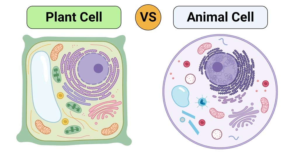
Plant Cell Vs Animal Cell Definition 25 Differences With Cell Organelles
Organelles shared by both plant animal cells.

Organelles in both plant and animal cells. They are jelly-like substances found between the cell membrane and nucleus. Vacuoles are storage sacs for solid or liquid contents. 7 rows Animal cells and plant cells also contain tiny objects called mitochondria in their cytoplasm.
Are they Plant animal or both plant and animal cells. Cell membrane Chromosomes Cytoplasm Mitochondria The plant and animal cells are eukaryotic and contain well developed cellular organelles. The central vacuole of some plant cells may occupy 50-90 of the cell volume.
Their primary function is to -. Golgi apparatus and ribosomes exist in both plant and animal cells. Cell organelle that releases energy in a process called cellular respiration.
While both plant and animal cells are eukaryotic and share many similarities they also differ in several ways. Vacuoles are round organelles found in both plant and animal cells. Discuss these differences in relation to the activities of plants and animals.
Amoeba acquires its food by the process of 30. Through how many membranes would a molecule have to pass in going from the interior of a chloroplast to the interior of a mitochondrion. The function of the mitochondria is to break down sugar molecules into ATP energy for both plant and animal cells.
The cytoplasm is one of the essential components of the cell where all the cell organelles are embedded. Vacuoles in animal cells are much smaller. Learn about the key differences between these two cell types in this lesson.
The cell wall and chloroplast are. As stated above both plant and animal cells share a few common cell organelles as. Learn with flashcards games and more for free.
In plant cells vacuoles are full of cell sap and provide turgidity swollen and distended or congested and rigidity to the cell. Rough Smooth Endoplasmic Recticulum. The mitochondria is located in the cytoplasm but outside of the nucleus.
Centrioles exist only in animals but not in plant cells. Secondly what are the main differences between plant and animal cells. Infoldings of the inner membrane of a mitochondrion.
Learn the following terms and their definitions. Structurally plant and animal cells are very similar because they are both eukaryotic cells. Common organelles of plant animal cells are as follow.
They are mainly composed of water organic and inorganic compounds. The cytoplasm is present both in plant and animal cells. In animal but not plant cells.
They both contain membrane-bound organelles such as the nucleus mitochondria endoplasmic reticulum golgi apparatus lysosomes and peroxisomes. Large vacuoles are common in plant cells. The cell membrane cytoplasm chromosomes and mitochondria are the structures that are present in both the plant and.
37 Animal Cell Under Electron Microscope
We all do not forget that the human physique is quite problematic and a method I. The cell membrane also known as plasma membrane or plasmalemma consists of three layers when viewed under the electron microscope.

Y4uq Xpinhgurm
Microscopically animal cells from the same tissue of an animal will have varied sizes and shapes due to the lack of a rigid cell wall.
Animal cell under electron microscope. Animal Cell as shown above. IB Biology HLSLA Worksheet Microscopes and Electron micrographs Prokaryotic and Eukaryotic cells. It has small vacuoles.
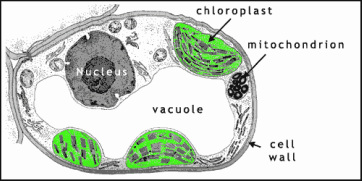
6 rows Structure of plant and animal cells under an electron microscope. Some of the cell organelles that can be observed under the light microscope include the cell wall cell membrane cytoplasm nucleus vacuole and chloroplasts. It is an electron micrograph of cells largest and most important organelle the mitochondria and is characterized by the following features Fig.
The Cell as Seen under the Electron Microscope. Diagram Of Animal Cell Under Electron Microscope Labeled. Table D leads to images of electron microscopes or protocols for tissue preparation.
1 The name mitochondria was given by Benda 1898 and their ma n function was brought to light by Kingsbury 1912. Monday April 5th 2021. This atlas offers informative texts on a lot of cell organelles and ultrastructures with detailed information in a generally intelligible way.
It is completed by a vocabulary of microscopic anatomy. In the given figure of an animal cell as observed under an electron microscope. So lets begin by drawing a rough-oval shape.
2 Each mitochondria in section appears as sausage or cup or bowl shaped structure lined by double. Also know what does a animal cell look like under a microscope. But at the same time it is interpretive.
The three layers are composed of one layer of phospholipid sandwiched between two protein layers. The cell membrane is. Animal cells have a basic structure.
View under scanning electron microscope yeast cells of the. Under the intense radiation of the electron microscope 011 electron per Å 2 the question of viability of cells naturally arises because the amount of radiation absorbed during highmagnification imaging is sufficient to cause cell death. Typical Animal Cell With Labels Removed 3.
The ultrastructure of budding and dimorphic yeast cells observed with a scanning electron microscope sem and a transmission electron microscope tem after thin sectioning and freeze etching are then described followed by discussion of the regeneration of the cell wall ofcandida albicans. Below the basic structure is shown in the same animal cell on the left viewed with the light microscope and on the right with the transmission electron. Beneath a plant cells cell wall is a cell membrane.
Use your ebook to answer the questions below. Plant cell as shown above. Diagram Of Animal Cell Under Electron Microscope.
Most cells both animal and plant range in size between 1 and 100 micrometers and are thus visible only with the aid of a microscope. How is it different from animal cell. Asked Nov 28 2017 in Class IX Science by ashu Premium 930 points.
Typical Animal Cell Pinocytotic vesicle Lysosome Golgi vesicles Golgi vesicles rough ER endoplasmic reticulum Smooth ER no ribosomes Cell plasma membrane Mitochondrion Golgi apparatus Nucleolus Nucleus Centrioles 2 Each composed of 9 microtubule triplets Microtubules Cytoplasm Ribosome 2. Its a thin slice. These cell organelles perform specific functions within the cell.
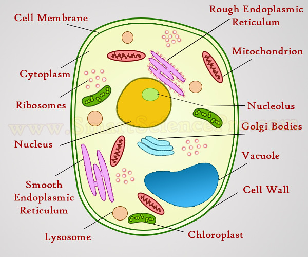
Heres a diagram of a plant cell. The diagram is very clear and labeled. Summarize two advantages and disadvantages of light microscopes.
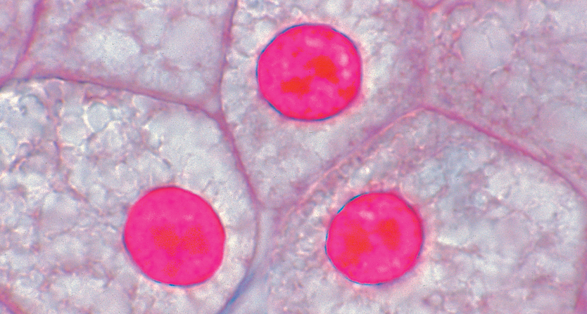
Structure of plant and animal. Human cheek cells are made of simple squamous epithelial cells which are flat cells with a round visible nucleus that cover the inside lining of the cheekC. Please show the substitute teacher your completed work as soon as you finish.
Illustrate only a plant cell as seen under electron microscope. However no obvious structural damage was apparent and several repeated scans gave the same images. Under a microscope plant cells from the same source will have a uniform size and shape.
It is flexible and has pores. Cell Structure and Function Student Hadi Yaafar Date May 24 2021 Instructions Please work independently. This feature was lost in the distant past by the single-celled organisms that gave rise to the kingdom Animalia.
Unlike the eukaryotic cells of plants and fungi animal cells do not have a cell wall. I Name the parts labelled as 1 to 10.
35+ Animal Cell Under Microscope
Similarly what does a animal cell look like under a microscope. Viewing animal cells under the microscope and calculating magnification.
Slide Animal Cell Sec
Likewise can rough endoplasmic reticulum be seen under a light microscope.
Animal cell under microscope. Under a microscope plant cells from the same source will have a uniform size and shape. Thousands of new high-quality pictures added every day. Animal cells under microscope animal cells under microscope labeled animal cells under microscope video animal cell under microscope 400x animal cell under microscope 40x animal cell under microscope 100x animal cell under microscope 10x animal cell.
Human cheek cells are made of simple squamous epithelial cells which are flat cells with a round visible nucleus that cover the inside lining of the cheekC. Under a microscope plant cells from the same source will have a uniform size and shape. These cell organelles perform specific functions within the cell.
The Cell as Seen under the Electron Microscope. Some of the cell organelles that can be observed under the light microscope include the cell wall cell membrane cytoplasm nucleus vacuole and chloroplasts. Section of beef marrow showing motor neurons in the anterior horn seen under a microscope.
Browse 163 animal cells under microscope stock photos and images available or start a new search to explore more stock photos and images. An animal cell also contains a cell membrane to keep all the organelles and cytoplasm contained but it lacks a cell. Microscopically animal cells from the same tissue of an animal will have varied sizes and shapes due to the lack of a rigid cell wall.
Observing Animal Plant Cells You will be observing plant and animal cells under the microscope. Scientists work hard to make accurate observations. Beneath a plant cells cell wall is a cell membrane.
To look at a cell close up we need a microscope. Plant cell as shown above. The shape of both cells were easily seen and some similarities and differences were.
Under the microscope animal cells appear different based on the type of the cell. Animal cell under the microscope A typical animal cell is 1020 μm in diameter which is about one-fifth the size of the smallest particle visible to the naked eye. Microscope is used extensively in cell biology microbiology biotechnology microelectronics nanophysics pharmacology mineralogy and forensics.
Cells are the smallest part of a living organism and are around 001 mm - 003 mm long. Beneath a plant cells cell wall is a cell membrane. In a plant cell the nucleus cell wall cell membrane and the cytoplasm were visible through a light microscope.
Diagrams from enchanted learning. The lesson will also take you through some exam questions on finding magnification using a scale and an image of a cell. The cell membrane is the outer most part of the cell which.
It is also used for medical diagnosis particularly while dealing with tissues or in smear tests on free cells or tissue fragments. Careful observations help scientists make predictions draw conclusions and find comparisons. Find animal cell under microscope stock images in HD and millions of other royalty-free stock photos illustrations and vectors in the Shutterstock collection.
Start studying Cells Under a Microscope Animal Cell Plant Cells. If a plant cell and an animal cell are observed under a microscope what are the characteristics of the cells that enable you to identify the cell as a plant cells. Animal Cell as shown above.
Viewing Animal Cells under a microscope. It is very important that you take the time to make careful observations when looking at cells. Find out how to observe cells under a microscope Click to.
In the animal cell the nucleus cell membrane and cytoplasm were visible through a light microscope. Learn vocabulary terms and more with flashcards games and other study tools. In this lesson you will learn the method of how to view an animal cell under the microscope.
Cell nuclei of a sea urchin egg Parechinidae seen under a microscope. In plant cells but not animal cells you will find i a cell wall ii green chloroplasts and iii vacuoles. Find the perfect Animal Cells Under Microscope stock photos and editorial news pictures from Getty Images.
Select from premium Animal Cells Under Microscope of the highest quality.
